Saturday, January 21, 2012

AP bio: Three Beneficial Bacteria

aA microscopic view of staphlyococcus epidermidis.  (The picture was taken from this site: http://www.flickr.com/photos/ajc1/2865663672/.)


Staphlyococcus Epidermidis


This bacteria is one of the many kinds of bacteria that live on the human skin. It is the first line of defense against harmful bacteria.  Most of these bacteria live in the hair follicles and go to the outer skin layer after humans wash their hands, but there are some that already live on the outermost skin layer.  They also live in the throat's mucous membranes.


A microscopic view of Lactobacilli in the vagina.  (This picture was taken from this site: http://textbookofbacteriology.net/normalflora.html.)


Lactobacilli


These bacteria are found in the vagina soon after a female child is born.  They make the vagina acidic as they make acid from glucose that was stored.  They stay there until other bacteria live in the vagina and it is not acidic anymore.  But when a female reaches puberty, the vagina is acidic again because a lot of Lactobacilli have inhabited it.  The Lactobacilli help protect the vagina from being attacked by other harmful bacteria and microorganisms.


A microscopic view of the viridans streptococcus in the throat.  This picture was taken from this site: http://en.wikipedia.org/wiki/Streptococcus_viridans.)

Viridans Streptococcus


These bacteria is may be passed on to a baby when it is going through the mother's birth canal.  Afterwards, the Viridans streptococcus is the main microbes that inhabit the mouth and the throat.  It stay like this for the rest of the person's life.  These bacteria are beneficial as they protect the mouth and throat when harmful bacteria or microorganisms try to invade.


AP bio: Factors Affecting Cell's Rate of Diffusion

(Based on the simulation on this site: http://www.mhhe.com/biosci/genbio/biolink/j_explorations/ch02expl.htm.)

When the simulation begins, the cell has a surface area of 3 and a relative diffusion rate of 1.  When I increased the villi percent of the cell surface area, there was no effect on the relative diffusion rate.  When I cell and the dimple percent of the cell surface area, there was no effect either.  However, when I increased the radius from 1X to 10X, the relative diffusion rate decreased to 0.10..  Also, when I increased the cell shape from a ratio of 1:1 to 3:1, the relative diffusion rate increased to 2.08.  Therefore, it is supported that an increase in radius means a decrease in a cell's rate of diffusion and an increase in the cell shape ratio means an increase in the cell's rate of diffusion.

AP Bio: Cellular Respiration VS Photosynthesis

A simplified version of the process of photosynthesis.  (This picture was taken from this site:  http://mrskingsbioweb.com/Biology.html.)


Photosynthesis
6CO2 + 6H2O + energy -> 6O2+ C6H12O6
Photosynthesis is the process that converts the energy from sunlight to the glucose, which is stored as organic molecules.  This is a process that takes place inside photosynthetic organisms.  These autotrophs, or self-feeding organisms, use the process to convert light energy/sunlight into glucose (sugar) to use for food/energy.  The process of photosynthesis includes the light dependent reactions, the Calvin Cycle (dark reactions), and the electron transport chain (ETC).  The light dependent reactions and the ETC (the first part of photosynthesis) occurs in the thylakoid membranes, water and light is absorbed.  NADPH provides the energy for the reaction to occur.  Oxygen, ATP, and NADPH is produced.  In the second part of photosynthesis (the Calvin cycle), ATP, NADPH, and CO2 is used to produce sugars and NADP+.  Th NADP+ is reused in the light dependent reactions.


A simplified version of the process of cellular respiration.  (This picture was taken from this site: http://www.hyperbaric-oxygen-info.com/aerobic-cellular-respiration.html.)


Cellular Respiration
6O2 + C6H12O6 --> 6H2O + 6CO2 + energy
Unlike photosynthesis, cellular respiration converts the stored organic molecules of glucose into energy.  It includes glycolysis, the Krebs Cycle, and the electron transport chain (ETC).  in glycolysis, the reactants are glucose and the products are some ATP electrons and pyruvic acid.  This occurs in teh cell's cytoplasm.  The pyruvic acid is sent to the mitochondria, where it goes through the Kerbs cycle.  More ATP is formed, and electrons carried the NADH and FADH2 are sent to the ETC.  The ETC is in a mitochondrial membrane and has a mechanism called chemiosmosis.  At the end of the ETC, a hydrogen gradient is created by proton-motive force as the hydrogen ions move across the mitochondrial membrane into the cell and a phosphate is added to adenine diphosphate to produce adenine triphosphate (ATP).  ATP provides for most of the energy used by the cell.


Source: Campbell and Reece's Biology, Sixth Edition

Thursday, January 12, 2012

Anatomy and Physiology: Skull and Bone Differences Based on Gender and Race

male and female skull differences (this picture was taken fromhttp://squinting-at-bones.blogspot.com/2011_07_01_archive.html)


Gender
Male skulls have a robust occipital protuberance, larger and more downward protruding mastoid process, zygomatic ridge extends beyond external auditory meatus, larger more distinct temporal line, and pronounced superciliary arches.

Female Skulls have vertical and rounded frontal, zygomatic doesn't extend beyond external auditory meatus, smaller less downward projecting mastoid process, and parietal bossing.

Race
Caucasoids skulls have narrow pointed mastoid process, rounded/elongated oval sagittal outline, small degree of facial prognathism, high and peaked nasal cross section, and a projecting chin.

American Black skulls have oblique mastoid process, high degree of facial prognathism, low and broad nasal cross section, and a elongated sagittal outline.

Asian skulls have a flatter frontal lobe, low degree of facial prognathism, a small and low nasal cross section, a less projecting chin, and a more rounded mastoid process.

Thursday, November 10, 2011

Anatomy and Physiology: Dead Men Do Tell Tales

(This picture was taken from this site: http://www.42ndpage.com/science/).

One thing I found very interesting in William R. Maples' Dead Men Do Tell Tales is the large amount of information a bone can tell about the dead person if you know how to look at it.  For example, the author and a professor used a skull that was found in a lake and tied to a pole to figure out that the skull was a World War II trophy.  Using the scorch marks and other features on the skull, they were able to determine that the skull belonged to a Japanese man who was probably a soldier killed during WWII and his skull was taken by an American soldier as a sort of prize.  The soldier got tired of it, but needed a place to dispose of the skull so he threw the skull in a lake and tied it to a pole for good measure.  And all this was from a single skull!

Source: William R. Maples' Dead Men Do Tell Tales

Saturday, November 5, 2011

Anatomy and Physiology: Two Ways of Bone Growth and Development

Bones can develop embryonically or postembryonically.  Bones use hyaline cartilage structures as "models" during bone formation or ossification.  The picture below shows the process of bone growth, both embryonic and postembryonic.

(This picture is taken from this site: http://training.seer.cancer.gov/anatomy/skeletal/growth.html).


During the embryonic development of the bone, the hyaline cartilage model is covered by oseoblasts (bone-forming cells).  The cartilage model would remain there until it is digested away and a medullary cavity opens up within the new bone.

During postembryonic development (bone development that happens after birth), most hyaline cartilage models are replaced by bone except the articular cartilages, which cover the bone ends, and the epiphyseal plates. The articular cartilages are never converted into bone because they reduce friction at the joints.  During long bone growth after birth, the long bones lengthen and widen.  When a long bone lengthens, new cartilage is always being formed on external side of the articular cartilage and the epiphyseal plate while the old cartilage is broken down and replaced by bony matrix on the internal side of the articular cartilage and medullary cavity.  When a long bone widens, it undergoes appositional growth, in which the osteoblasts in the periosteum add bone tissue to the outer surface of the diaphysis (shaft of the bone) while the osteoclasts (bone-destroying) cells break down bone tissue in the inner surface of the diaphysis.

Cartilage and bone tissue is constantly being replaced during  a bone's appositional growth. (This picture is taken from this site: http://www.rci.rutgers.edu/~uzwiak/AnatPhys/APFallLect8.html).
Sources:
Elaine N. Marieb's Essentials of Human Anatomy & Physiology
Dawn A. Tamarkin's website http://faculty.stcc.edu/AandP/AP/AP1pages/Units5to9/bone/bonedev.htm

Thursday, October 20, 2011

AP bio: C3. C4, and CAM Plants


(Dandelions are example of plants that use the C3 pathway.  This picture is taken from this site: http://www.google.com/imgres?q=dandelion&num=10&hl=en&gbv=2&biw=1024&bih=610&tbm=isch&tbnid=ZGn9W6PPMOALHM:&imgrefurl=http://www.manataka.org/page1126.html&docid=766naOQmHrWzVM&imgurl=http://www.manataka.org/images/dandelion.jpg&w=425&h=319&ei=eTCqTrLNBdLQiALO5_GuCw&zoom=1&iact=rc&dur=249&sig=110919301459896358943&sqi=2&page=1&tbnh=115&tbnw=153&start=0&ndsp=15&ved=1t:429,r:5,s:0&tx=80&ty=74)
C3 Plants: When it is hot or dry, C3 plants close their stomata to prevent water loss.  Because of this, photorespiration occurs when the carbon dioxide is less concentrated in the air spaces in the leaf, which slows down the Calvin Cycle.  Oxygen and carbon dioxide accumulate in the leaf and rubisco adds oxygen to the RuBP.  The product is broken down and a two-carbon compound is broken down, releasing carbon dioxide.  Cactus is an example of C3 plants.


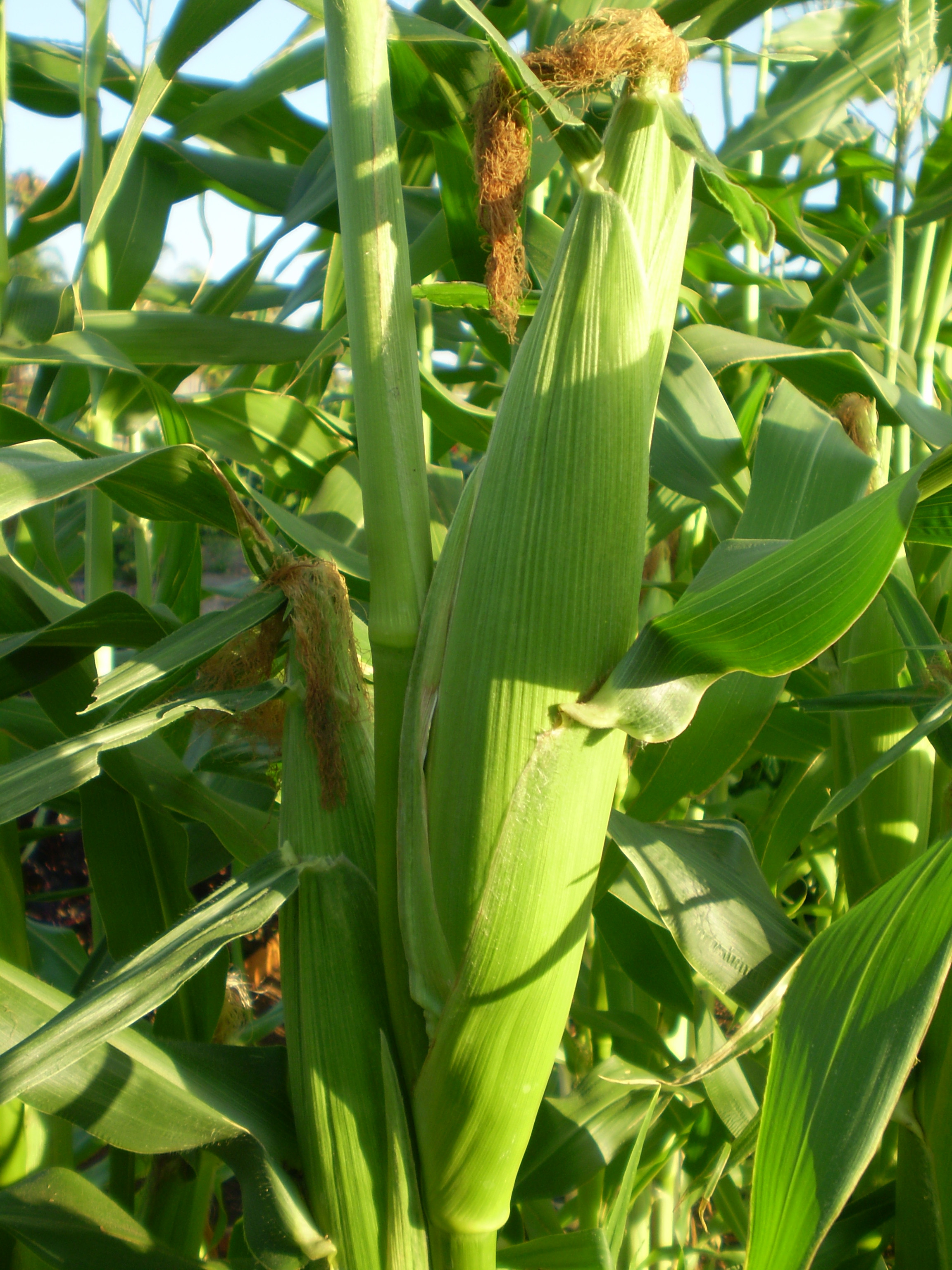
(Corn is an example of a plant that uses the C4 pathway.  This picture is taken from this site: http://www.google.com/imgres?q=c4+plants+examples&num=10&um=1&hl=en&biw=1024&bih=610&tbm=isch&tbnid=7WCZi_3MfDP0FM:&imgrefurl=http://fhs-bio-wiki.pbworks.com/w/page/12145744/CAM%2520and%2520C4%2520plants&docid=dXcRfJ_0a06RsM&imgurl=http://fhs-bio-wiki.pbworks.com/w/page/12145744/f/corn.jpg&w=2304&h=3072&ei=PS6qTtbtA4riiALW-uiYCw&zoom=1&iact=hc&vpx=424&vpy=114&dur=2383&hovh=259&hovw=194&tx=35&ty=276&sig=110919301459896358943&sqi=2&page=1&tbnh=128&tbnw=93&start=0&ndsp=15&ved=1t:429,r:2,s:0)
C4 Plants: Before the Calvin Cycle begins, PEP carboxylase (an enzyme) helps add carbon dioxide is to PEP (a 3-carbon compound).  The product is a four-carbon compound that is made in the leaf's mesophyll cells and is transported to the bundle-sheath cells (located around veins of the leaf).  The four-carbon compound is broken down, releasing carbon dioxide and initiating the Calvin Cycle.


(Pineapples are an example of plants that use the CAM pathway.  This picture is taken from this site: http://www.google.com/imgres?q=pineapple+tree&hl=en&gbv=2&biw=1024&bih=610&tbm=isch&tbnid=yrwJi6IcL45PpM:&imgrefurl=http://www.mrbigben.com/food/pineapple.html&docid=OvrVOWQ13DdWwM&imgurl=http://www.mrbigben.com/food/twin-fruit-pineapple-719390.JPG&w=640&h=480&ei=KS-qTtvAGse0iQLB66G1Cw&zoom=1&iact=hc&vpx=526&vpy=177&dur=2051&hovh=194&hovw=259&tx=158&ty=214&sig=110919301459896358943&page=1&tbnh=118&tbnw=153&start=0&ndsp=20&ved=1t:429,r:4,s:0)
CAM Plants: These plants may also close their stomata to prevent water loss and beak down compounds to release carbon dioxide during the day.  However, the crassulacean acid metabolism (CAM) pathway doesn't separate the process of carbon fixation from the Calvin Cycle, unlike the C4 pathway.

Source: Campbell and Reece's Biology, Sixth Edition