Saturday, January 21, 2012

AP bio: Phyla of Protozoa

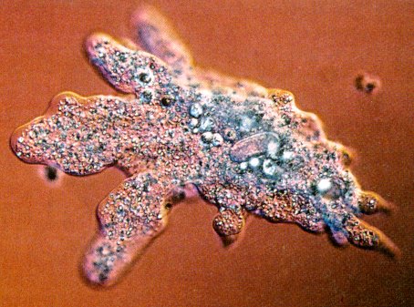
A microscopic view of a amoeba, a protist from the phylum Sarcomastigophora.  This picture was from: http://homosapienssaveyourearth.blogspot.com/2012/01/it-2-01-amoebas-heavenly-dream.html.)


Phylum Sarcomastigophora
The protozoa in this phyla can be unicellular or colonial.  They can move around by flagella, pseudopodia, or both, and usually reproduce through binary fission and sexual reproduction.  An example of a protozoa from this phylum include the amoeba, which uses a psueopod or "fake foot" to move around.


A microscopic view of the parasitic protozoan labyrinthuloides haliotidis, which is from the phylum labryinthormorpha.  This picture is from: http://eol.org/pages/21984/overview.)


Phylum Labryinthormorpha
The protists in this phylum are spindle-shaped or spherical.  They move by gliding on mucous.  Most of these protozoans are marine and are parasitic towards algae.  An example is labyrinthuloides haliotidis, which is a parasite for abalone.

A microscopic view of mature Plasmodium-malariae.  This picture is from: http://en.wikipedia.org/wiki/Plasmodium_malariae.


Phylum Apicomplexa
These protozoans have a stage in their life cycle where they produce spores.  They have a unique structure called the apical complex (a arrangement of vacuoles, fibers, and other organelles at one end of the cell).  They have complex life cycle in which they inhabit two hosts (ex: a mammal and a mosquito).  The life cycle has an alternation of generations of haploid and diploid and they can reproduce asexually or sexually.  At some point in life, they produce a lot of small infectious organisms through a process called schizogony.  An example of this kind of protozoan include the pathogen Plasmodium-malariae.


This shows a host cell that is infected by Brachiola algerae, a protozoan from the phylum microspora.  This picture is from: http://www.cns.fr/spip/Brachiola-algerae-a-multi-host.html.


Phylum Microspora
These protozoans are intracellular parasites.  They do not have mitochondria and some are pathogens of insects, and are transmitted by a spore.  Five kinds have been seen in the implications of human diseases, such as in people with AIDS.  An example is the Brachiola algerae, which is a pathogen transmitted by mosquitoes. 


A microscopic view of Bonamia ostreae infecting oysters.  This picture is from: http://www.scotland.gov.uk/Topics/marine/Fish-Shellfish/18364/18610/diseases/notifiableDisease/Bonamiaostreae.


Phylum Ascetospora
These protozoans are parasitic protists.  They have spores for reproduction and do not have polar filaments or polar caps, and are parasitic in mollusks.  An example is the Bonamia ostreae, which infects the flat oyster Ostrea edulis (a type of mollusk).  


The spores of Kudoa thyrsites, a parasitic protist from the phylum Myxozoa.  This picture is from: http://www.journalofparasitology.org/doi/abs/10.1645/GE-548R.1.)


Phylum Myxozoa
These protozoans are also parasitic protists.  Like the phylum Ascetospora, they also have resistant spores.  The spres have one to six polar filaments and these protists are parasitic in fish, both freshwater and marine.  They can cause economic problems in salmon that the farmed.  An example is kudoa thyrsites, which attacks fish muscle and loosens it.


A microscopic view of Paramecium caudatum, a protist from the phylum Ciliophora.  This picture is from: http://101science.com/paramecium.htm.)


Phylum Ciliophora
Phylum Ciliophora is the largest out of the phyla of protozoa.  These protozoans are characterized by using cilia to move around.  They have various unique structures, such as tentacles or toxicysts (threadlike darts).  To feed, they capture food with the cilia and the food is eventually passed to the vacuoles to digest.  These protozoans can reproduce asexually by binary fission and sexually by conjugation.  Most of them are harmless but some are parasites.  An example is the Paramecium caudatum, a harmless, free-living protist. 


Sources:
http://highered.mcgraw-hill.com/sites/0072320419/student_view0/chapter27/study_outline.html
Campbell and Reece's Biology, Sixth Edition

AP bio: Comparing Bacteria, Virus, Prion, and Protist

A typhoid bacterium.  (This picture is from: http://news.softpedia.com/news/Typhoid-Bacterium-Accompanied-Us-Along-Our-Evolution-41046.shtml.)


Bacteria
Bacteria can adapt very well to environment that change constantly because they have short generation spans.  During these short generation spans, they can undergo genetic recombination.  This way, so when they reproduce through various ways such as transformation, transduction, and conjugation, the new DNA will replicate along with their own original DNA.  These new, foreign DNA are expressed in the new strains of transformed bacteria.  This also lets each bacterium adapt their metabolic processes with the changes of their environment.

A diagram of the structure of a vius.  (This picture was from: http://www.armageddononline.org/viruses.html.)


Virus
Unlike bacteria, virus are not actually alive because they cannot survive outside a host.  They are basically just genes in a protective coat.  The parts of a virus includes its capsid, protein coat, viral envelope, and nucleic acid.  Bacteriophages are the group of viruses that can infect bacteria.  They reproduce through the lytic and lysogenic cycles.  In the lytic cycle, they attack the host cell and when the cell bursts, the new viruses move on to destroy other host cells.  In the lysogenic cycle, the virus may lie in wait in the host cell for a long time before the lytic cycle is activated and it destroys the host cell.

The structure of a prion.  (This picture was from: http://www.physorg.com/news153681509.html.)


Prion
Prions are simple infectious agents.  They are basically protein that are misfolded.  It has no genetic material.  Although they are proteins, they can reproduce on their own and become infectious agents.  In humans, they may have been linked with the Creutzfeldt-Jakob Disease (CJD) and the Gerstmann-Straussler-Scheinker Syndrome.

A microscopic view of a paramecium, a protist.  (This picture was from: http://www.mlms.loganschools.org/~ckircalli/homework/MATH_SCIENCE%20LINK%20PAGES/PROTIST%20INTERNET%20LESSON.html.)


Protist
Protists are eukaryotic microoorganisms.  They are classified in the Protista kingdom, which also includes unicellular organisms.  Sometimes, their behavior might make them pathogens that infect plants and animals.  Protists have a lot of different ways of feeding, such as filter feeding in flagellates and endocytosis (usually phagocytosis or cell-eating, but sometime pinocytosis or cell-drinking.)  Some reproduce sexually with gametes or asexually through binary fission.  Other protists are more complicated and have life cycles in which some forms of the organism reproduce sexually and others asexually.

Anatomy and Physiology: Mink and Human Muscle Similarities

(This picture was from: http://uscunion.sc.edu/biology/Dissections/MinkMuscArm2L.jpg.)
Note the similarities in the structure of the mink and human arm muscles.  (This picture was from: http://anatomyeshs.wikispaces.com/Ch.8+Muscular+System.)


During the mink dissection, I saw that many of the muscles on the mink had the same shapes as their human counterparts.  For example, when we identified some of the muscles in the leg, the mink had the muscles biceps femoris, sartorius, gastrocnemius, adductor longus and femoris, semitendinosus, etc.  All of these muscles are in the human leg.  They are also similar in shape.  For instance, the biceps femoris in the mink and the human are pennate muscles, or muscle that attaches at a oblique angle to its tendon.  The thing that surprised me most about the mink was the numerous similarities in muscle structure and function.  It reminded me of the evidence that was used by evolutionists to back up Charles Darwin's theory of evolution; one of the evidence was similarities in bone and muscle structure.

Anatomy and Physiology: Mink and Human Muscle Shapes

The biceps brachii is an example of a fusiform muscle.  It is wider in the middle and tapers at the ends.  (This picture was from: http://athleticconditioning.blogspot.com/2010/05/building-bigger-biceps.html.)


Fusiform:
Fusiform muscles are spindle-shaped.  Examples include many of the arm muscles, such as the biceps brachii, flexor carpi radialis, extensor radialis longus, and brachioradialis.


Location of the quadratus femoris, a quadrate -shaped muscle.  (This picture was from: http://medicina.ronnie.cz/c-1451-svaly-kycelniho-kloubu.html.)


Quadrate:
Quadrate muscles are quadrilateral-shaped.  Examples include the pronator quadratus on the distal forearm and the quadratus femoris on the thigh.


Location of the external oblique muscle, a flat muscle with aponeurosis.  (This picture was from: http://www.weight4me.com/fitness/muscles/external_oblique.htm.)


Flat with Aponeurosis:
These muscles have layers of flat, broad tendons.  Examples include abdominal muscles, such as the external oblique muscle and the internal oblique muscle.


Location of the extensor digitorum longus, a unipennate muscle, in relation to the muscle around it.  (This picture was from: http://www.getbodysmart.com/ap/muscularsystem/footmuscles/menu/menu.html.)


Unipennate:
Unipennate muscles have muscle fibers that stretch in the same direction.  Examples include the extensor digitorum longus and the brachialis.


Location of the rectus femoris in relation to the other muscles on the femur.  (This picture was from: http://www.t-nation.com/free_online_article/sports_body_training_performance_repair/18_tips_for_bulletproof_knees.)


Bipennate:
Bipennate muscles have two rows of muscle fibers that stretch in opposite directions and have a central tendon.  Examples include the rectus femoris and the gastrocnemius.


Location of the deltoid muscles.  (This picture was from: http://www.sciencelearn.org.nz/Contexts/Sporting-Edge/Sci-Media/Images/Deltoid-muscles.)


Multipennate:
Multipennate muscles have more than two rows of muscle fibers that stretch in different directions and have a central tendon that branch off into two or more tendons.  Examples include the deltoid muscle and the pectoralis major muscle.


Source:
The list of muscle shapes was taken from this site: http://www.netterimages.com/image/7453.htm.)

AP bio: Extreme Organism - Tubeworms


This picture is taken from: http://www.nsf.gov/news/special_reports/sfs/popup/life_vc_tubeworms.htm.)


Tubeworms (Riftia Pachyptila) live in the hydrothermal vents on the Pacific Ocean floor.  Here, the conditions are harsh.  The liquid from the hot vents mixes with the cold seawater.  Tubeworms look like giant lipsticks and can grow to approximately 3 meters (8 feet) tall.  They live in white tubes that are made of chitin, a tough, natural material that is chemically related to cellulose.

The chemical structure of chitin.  (This picture was taken from: http://academic.brooklyn.cuny.edu/biology/bio4fv/page/chitin.html.)


These strange creatures do not have a mouth, eyes, or a stomach.  They have a symbiotic relationship with the bacteria that live inside them, which ensures their survival.  The bacteria convert the chemicals from the vents into food for the worms through the process of chemosynthesis.  The red part of the tubeworm ia where it breaths.  The blood in this part of the tubeworm has a special kinds of hemoglobin that has a very high chance of reacting with the oxygen in the seawater.

AP bio: Bacterial Transformation and Transduction

The different strains of foreign DNA expressed in E. coli in Cohen and Boyer's experiment. 
Putting the bacteria into heat shock to induce them to take in foreign DNA.
One of the foreign DNA from the plasmid that is expressed in the transformed E. coli bacteria that grew on the bacteria culture plate.


Transformation: In this process, bacteria take in DNA molecules.  When they reproduce, the DNA molecule will also replicate with their own DNA.  The bacteria can be induced to take in certain DNA molecules, such as in Stanley Cohen and Herbert Boyer's experiment.  Cohen and Boyer inserted recombinant DNA into E. coli bacteria via a plasmid.  They created a heat shock by lowering and raising the temperature to make the bacteria take in the plasmid, then put the transformed bacteria into a culture plate.  The result is that the transformed bacteria would express the foreign DNA.


The process of bacterial transduction.


Transduction: In this process, bacteria transfer foreign DNA to each other via a virus.  When bactiophages infect a bacterium, the viral DNA can be included in the bacterial genome.  This way, when the bacteria reproduces that viral DNA is also reproduced along with the bacteria's own original DNA.


Sources:
The pictures and information for transformation are taken from this site: http://www.dnalc.org/view/15916-DNA-transformation.html.
The information for the transduction is from Campbell and Reece's Biology, Sixth Edition.  The picture for transduction is from: http://bacteriakingdoms.com/transduction-bacteria/.

Anatomy and Physiology: Art and Anatomy

Joseph Nollekens' Minerva.  (This picture was taken from: http://www.all-art.org/Architecture/21-2.htm.)


Ancient Greece and Rome:  The art pieces from the time during ancient Greece and Rome looked perfect.  An example is the sculpture Minerva by Joseph Nollekens.  The body is portrayed in perfection; the people were beautiful, and the features of the face were beautiful.  The ancient Greeks were obsessed with perfection and this was reflected in their art, especially the paintings and sculptures of the gods and goddesses that they worshipped.


Realist painter Jean-Francois Millet's Man with a Hoe.  (This picture was taken from: http://www.paintinghere.com/painting/Man_with_a_hoe_6237.html.)


Realism: During the realist era, there was a spur in the advancement of science and the people portrayed in the art pieces became more realistic.  For example, the painting Man with a Hoe by Jean-Francois Millet, the man who is working outside looks like an actual person.  The study of anatomy allowed the artist to produce a more realistic painting with a better knowledge of the human body.  When drawing people, muscles became more defined and the proportions of the body became more correct.